When I wrote last month’s post, I didn’t expect it to be relevant this soon, yet you will see that it is a fitting introduction for this one! A few days ago at the beginning of June, while scrutinising Sentinel-2 images of the Loire river turbid plume, my PhD supervisor spotted red patches in the sea. See for yourself:
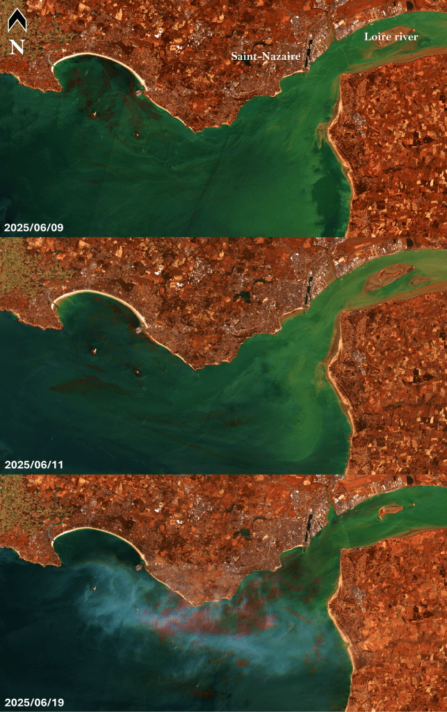
The mouth of the Loire river is located roughly 50 km west of Nantes, so we had a magnificent phytoplankton bloom right next door! And, given the location and the season, it was probably the most interesting kind of phytoplankton by far1: dinoflagellates!
In true oceanographer fashion, my colleagues and I set out to sea to get a closer look, and hopefully get some of these sea monsters back in the lab.
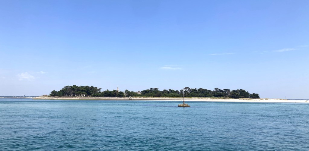
A beautiful day to sample some dinos
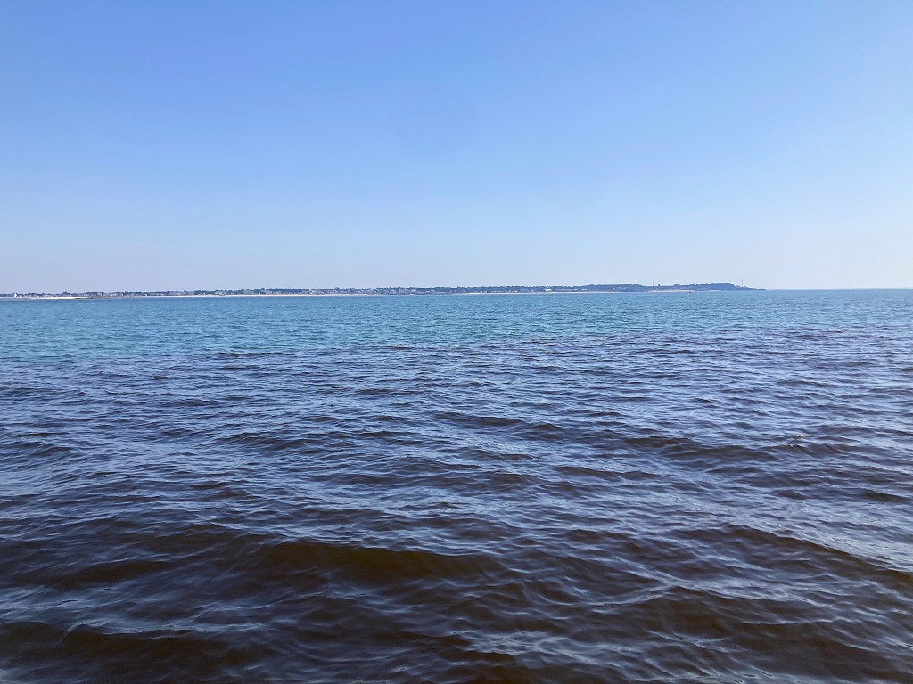
In the sunny morning of June 20th, we left the charming port of Le Croisic and went South then East to try to find some patches of coloured seawater. And patches we found! The water looked like black tea. Here is a photo where you can see the frontier between the patch and “normal” waters in the background, if you squint a little:
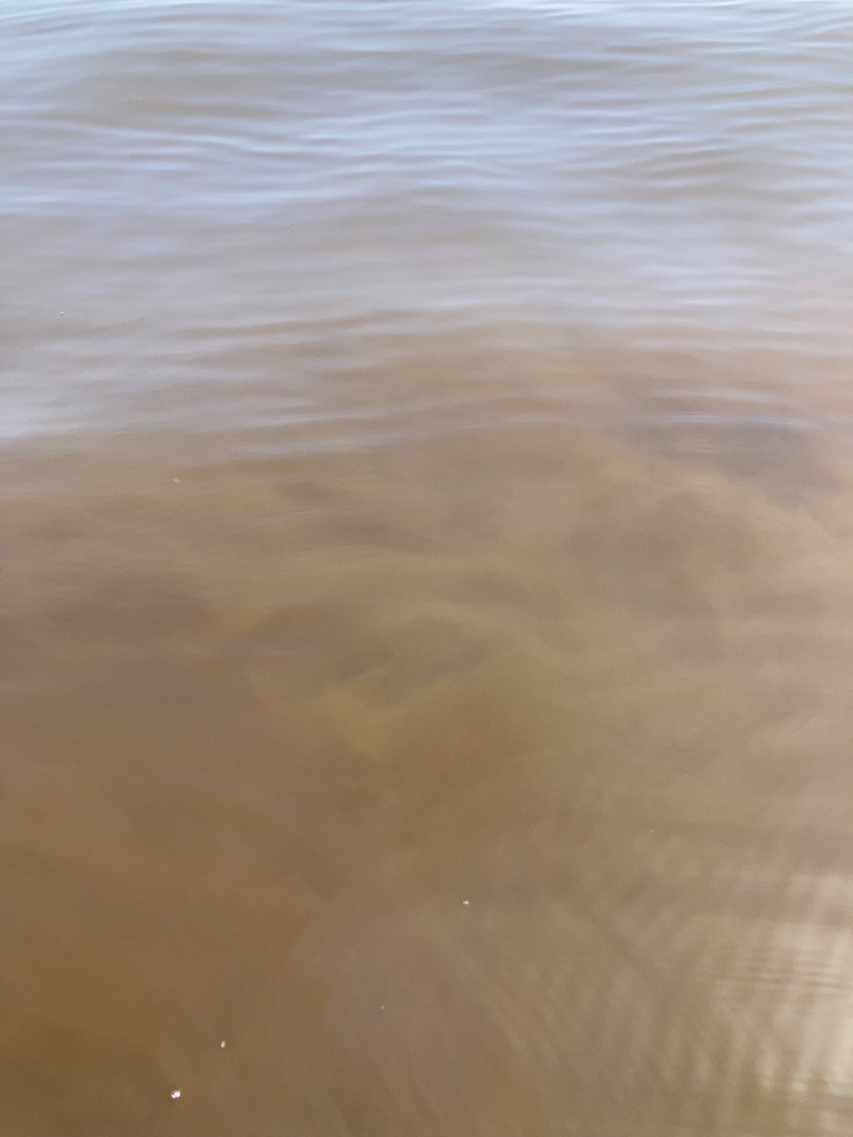
Directly next to the boat, in the more concentrated patches, we could distinguish swirls caused by myriads of cells drifting together:
After we finished sampling and had a little picnic on board, we headed back to the port, took a last gaze at the beautiful seaside, as I prepared to spend the rest of the day in the darkness of the microscopy lab.
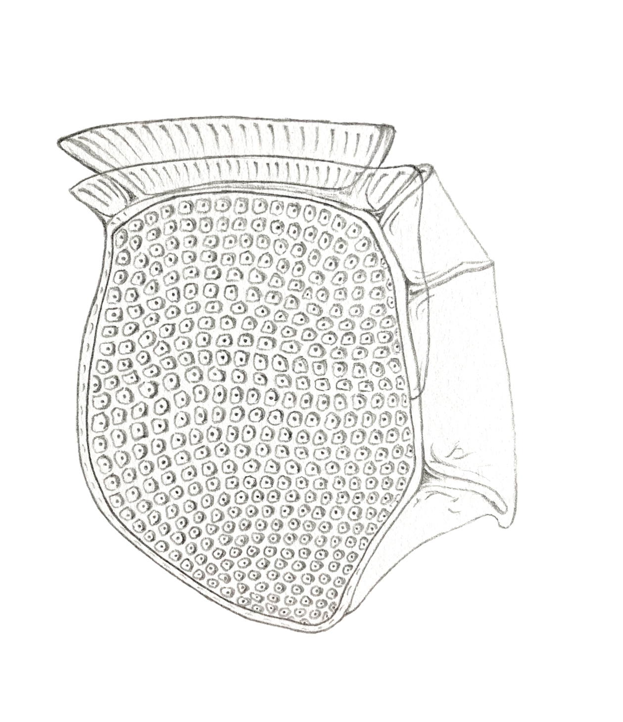
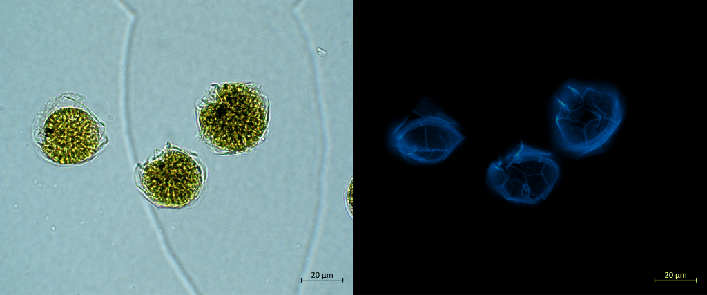
Alexandrium, you beauty
The cells of many dinoflagellate species are covered by a wall of cellulose called the theca. The ornamentation of the theca and the position of its different plates can help differentiate between species. After examination by an experienced dinoflagellate taxonomist in our team, we learned that our bloom was caused by Alexandrium tamarense2:
Beneath the theca is the heart of the cell, where the organelles lie and the magic of biology operates. After a drop of SYBR green and an excruciating 30-minute wait, I was ready to shoot some nuclei and chloroplasts! As so often with dinos, this microscopy session did not disappoint.
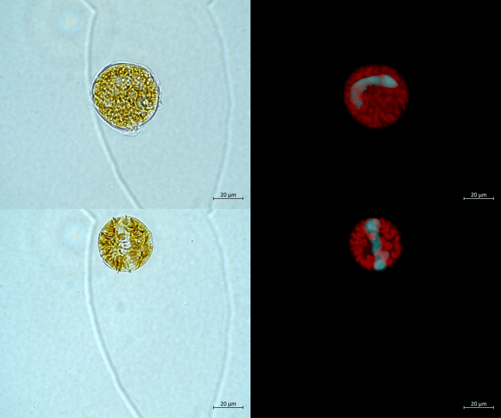
The cells are fully packed with chloroplasts, these little guys must have been doing some pretty good photosynthesis out there! They probably accumulated around the turbid plume because they grew on the nutrients brought by the river.
The nuclei are also quite a sight. They are horsehoe-shaped, which is not entirely unusual for dinos (see the photo of Prorocentrum micans in the very first post of this blog). Most fascinating: if you look very closely at the nucleus, you can see the individual chromosomes. In dinoflagellates they are permanently packed, an oddity compared to most other eukaryotes in which the chromatin is condensed only during cell division.
When the sea lights up at night
At 8 p.m., my institute closed for the week-end and I had to relunctantly leave the microscopy room, effectively ending one of the greatest workdays. Yet, I thought I could enjoy a bit more dinoflagellate mania before calling it a day. With some friends, we drove West for a dinner on the beach and waited for the night to settle. Arguably, the most mesmerizing feature of this Alexandrium bloom could only be witnessed well after the sunset.
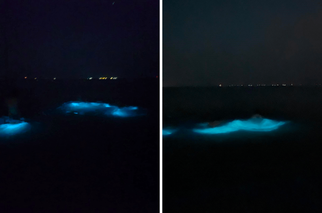
Every swimming stroke ignited ghostly blue flashes around our bodies. Billions of Alexandrium cells, disturbed by our movements, letting out bursts of bioluminescence. I don’t think calling this a magical moment is an overstatement, and I wish everybody has the chance to experience this in their lifetime.
Now, because this is a serious science blog, a few words about what’s happening here. Some dinoflagellates can emit bioluminescence at night, thanks to the well-known luminescent protein luciferin3. The emission of bioluminescence is triggered when the cell experiences shear stress, for example in breaking waves or when a clueless human is swimming nearby because it thinks biolumescence in the sea at night is bloody awesome. But more importantly, it activates when a copepod (a zooplanktonic predator of dinoflagellates) captures a cell and begins to manipulate it. The flash is so powerful that the copepod gets scared and rejects its prey, before quickly swimming away. This phenomenon has been described (and even filmed!) in another dinoflagellate species in this great article by Prevett and colleagues. Go check it out!
Honestly, the photos I’ve included do not do justice to the experience at all (and yet they are the best we could get!). It’s hard to describe the feeling, but I think you can get an idea if you imagine what it’s like being one of the dolphins in this video. Anyway, it was a fitting conclusion to what was already close to a perfect day.
See you next month!
The text and images (unless stated otherwise in the legend) in this blog post are under a Creative Commons Attribution license, which means that you can reuse them freely as long as you cite the author(s) properly.
- Personal opinion, obviously. ↩︎
- Which was a bit of a relief, as strains of this species are (so far) not toxic in our region, unlike their close cousin Alexandrium minutum! ↩︎
- I will not go into the details of what happens at the molecular and cellular levels, because this post is already long enoug and because I know too little on the subject. But this is a fascinating topic, that may deserve its own blog post some day! For now, if you can’t wait, you can read this article which seems quite detailed. ↩︎

Leave a comment