A curious case of shellfish poisoning in Japan
In the summer of 1976, coastal towns in Miyagi prefecture in northeastern Japan were faced with an unusual epidemic: 42 people presented symptoms of vomiting and diarrhea after they had eaten mussels.
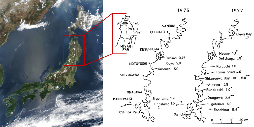
One year later, a similar outbreak occured, this time all along the eastern coast of the Tohoku region (see the map below). Some cases were recorded as far away as Tokyo, where poisonous mussels and scallops had been shipped. From the 3rd of June to the 22nd of July 1977, a total of 122 confirmed cases of shellfish poisoning were reported. Although all patients recovered within 3 days, this was undoubtedly a serious public health concern, so Takeshi Yasumoto and his colleagues from the Laboratory of Food Hygiene of Tohoku University began to investigate.
Bacterial contamination was quickly ruled out after microbial analysis of leftover meals and patients’ excreta (a nice word for vomit and/or poop) failed to detect any pathogenic bacteria. Some clues, such as the apparent seasonal periodicity, pointed towards a phytoplanktonic suspect. Forty years earlier, in 1937, American researchers (Sommer and Meyer) had described in detail a series of shellfish-related poisonings that had occurred during the previous decade along the Californian coast. People, sometimes dozens at a time, inexplicably fell ill after eating mussels. Victims reported tingling sensations and numbness in their lips, fingers and toes, followed by a paralysis of voluntary movements and, in the most extreme cases, death by respiratory failure.
Sommer, Meyer and their colleagues noticed a strange pattern: during poisoning outbreaks, a microscopic phytoplankton was present in unusually high numbers in seawater around the incriminated mussel beds: the dinoflagellate Gonyaulax catenella1. When they injected lab mice with extracts of G. catenella samples, the poor animals died just like their conspecifics that had received a dose of toxic shellfish slurry. They had identified the source of the poisonings: by filtering water full of the toxic G. catenella cells, mussels accumulated the toxin in their flesh, which made them so dangerous. Sommer and Meyer appropriately named the syndrome Paralytic Shellfish Poisoning (PSP).
Searching for the culprit
Back to the future, in 1977 Japan, Yasumoto and his team were puzzled. The whole situation reminded them furiously of PSP, but the symptoms did not match. After extracting the flesh of toxic shellfish into solvents of different polarity, they injected mice with the different fractions (water-soluble or fat-soluble)2. It became apparent that the toxic chemical was fat-soluble, contrary to the water-soluble saxitoxin, that causes PSP. The conclusion was evident: this was a new, unknown type of shellfish poisoning, and the identity of the toxic phytoplankton responsible for it was a mistery.
The culprit seemed particularly discreet. Toxic microalgal blooms often occur when one species proliferates to such an extent that it can be visible to the naked eye because the colour of the sea changes drastically, phenomena known as “red tides“. This was not the case here: no phytoplankton species was found in unusually high numbers at the time of poisoning outbreaks. Yasumoto and his team decided to take a patient and methodic approach: they would sample shellfish and plankton at the beginning of summer in a coastal bay, and try to find a correlation between a microalgal species’ abundance and the toxicity of shellfish.
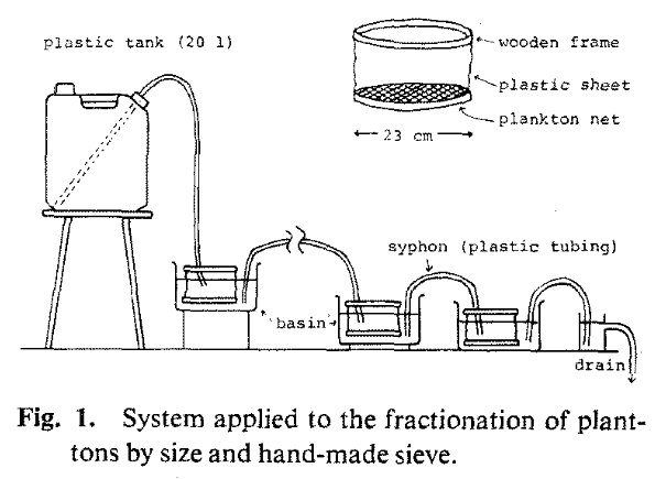
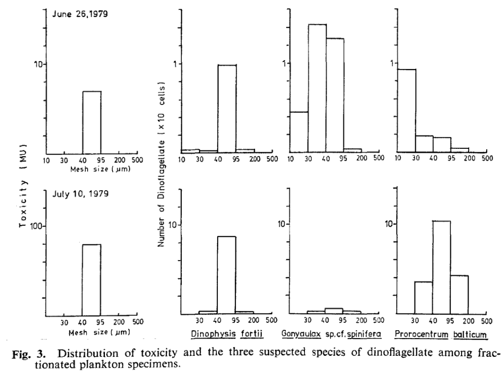
At the end of June 1979, their patience was rewarded: the mussels in their study site of Okkirai bay became toxic. With an ingenious system of serial filtration, they partitioned phytoplankton cells into different size fractions, and tested the toxicity of each fraction (once again, poor lab mice).
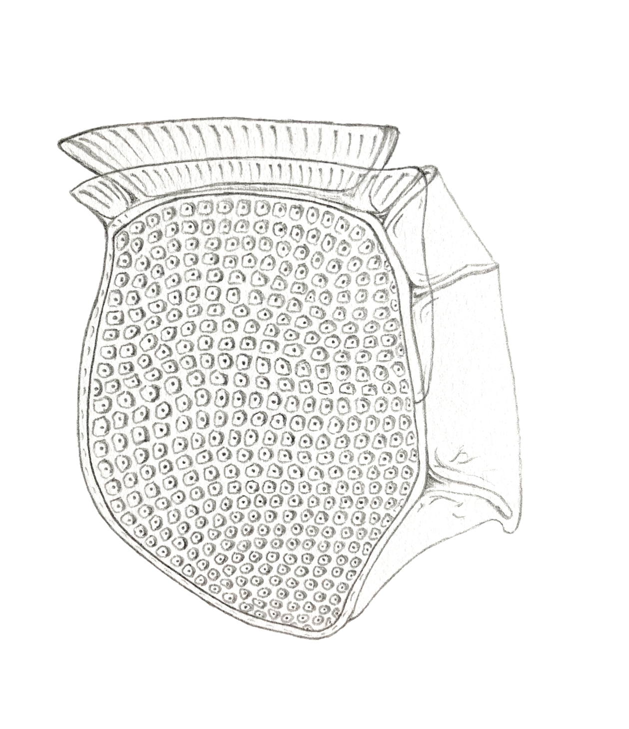
The results were quite clear: the 40-95 µm size class was the only toxic one, and the only phytoplankton species that was specifically contained in this fraction was the dinoflagellate Dinophysis fortii.
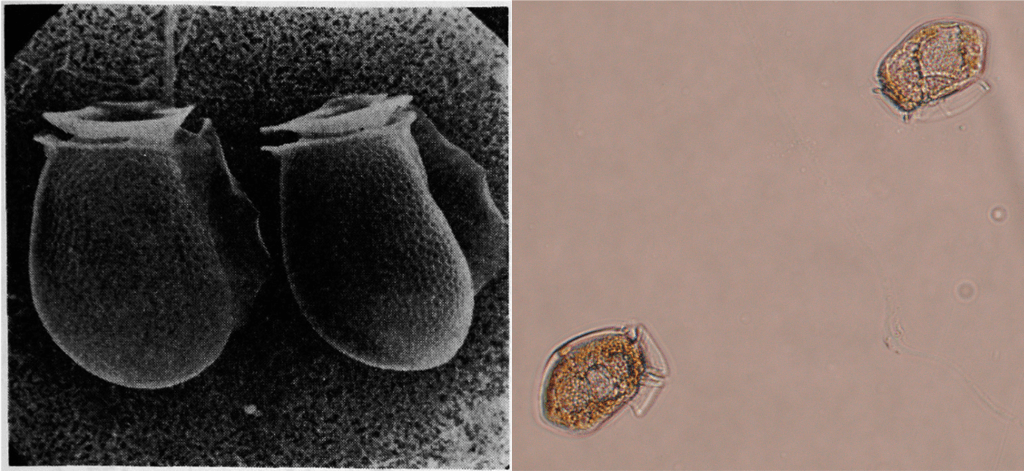
These results were confirmed in June 1980 when a new toxic episode occured: toxicity in phytoplankton samples was once again perfectly correlated with the abundance of D. fortii cells. The shellfish poisoning outbreaks in 1976 and 1977 had been caused by shellfish that had accumulated toxins by feeding on this dinoflagellate. Yasumoto and his colleagues published their findings in the Bulletin of the Japanese Society of Scientific Fisheries, naming the syndrome Diarrhetic Shellfish Poisoning (DSP). In their 1978 article on the description of the poisonings, Yasumoto and his colleagues noted:
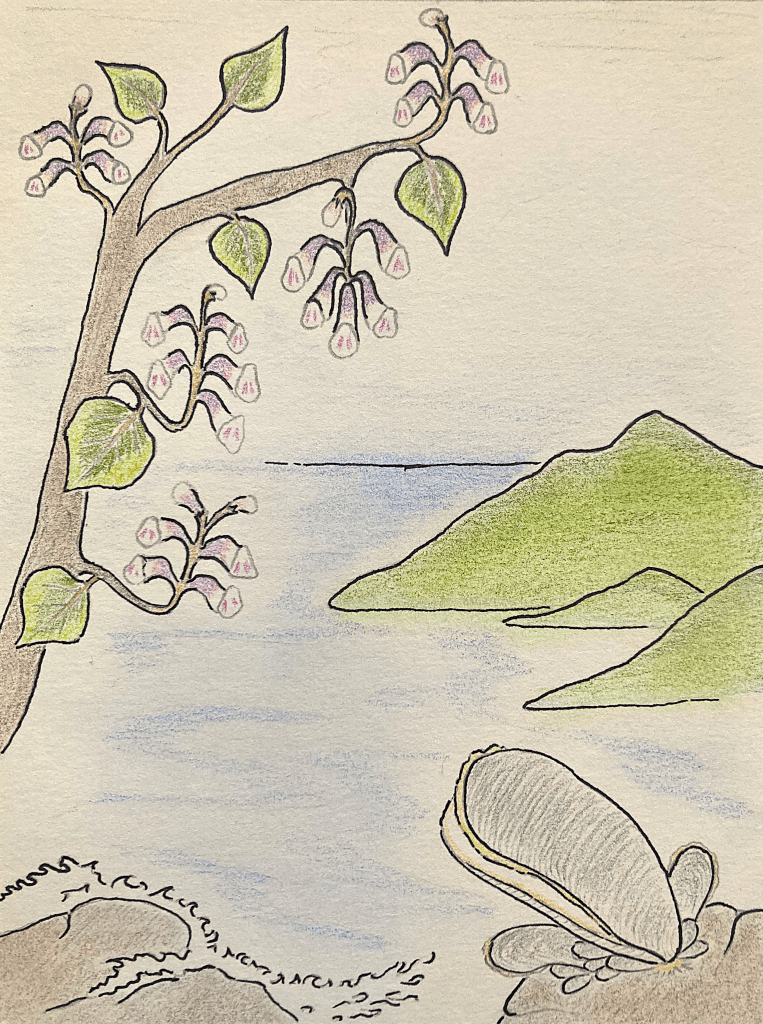
“Despite the lack of previous official record, however, such phenomenon seems to have existed since long as indicated by a widespread legend that mussels may be poisonous during the season of paulownia flowers“.
What a way to add some historical context AND a charming poetic touch!
Dinophysis goes global
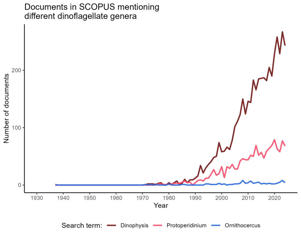
Two years later, in 1982, the same team published a chemical structure of the toxin responsible for DSP symptoms, that they named dinophysistoxin. In the following years, investigations outside Japan uncovered that other species in the Dinophysis genus were also producers of toxins, and were also associated with DSP events in the Netherlands and South Korea. The reveal that the widespread and relatively common dinoflagellate Dinophysis posed a serious threat to public health (and, indirectly, to the shellfish-farming industry) ignited the interest of the research community. As a proof: the number of documents in the scientific literature mentioning the term “Dinophysis” seems to have increased exponentially from the mid-1980s onwards, at a much higher rate than for non-toxic dinoflagellates3:
Research was much needed, as scientists would soon discover that toxicity was maybe not the strangest aspect of Dinophysis‘ biology…
This post is the first of what I think will be a 3-part series on Dinophysis. (Edit: the second part is now online! You can go read it here)
It is largely based on the 2 articles by Yasumoto et al. (1978 and 1980), and I hope it gives a fair presentation of their great work:
Yasumoto, T. et al. “Occurrence of a new type of shellfish poisoning in the Tohoku district.” Bulletin of the Japanese Society of Scientific Fisheries 44.11 (1978): 1249-1255.
Yasumoto, T. et al. “Identification of Dinophysis fortii as the causative organism of diarrhetic shellfish poisoning.” Bulletin of the Japanese Society of Scientific Fisheries 46.11 (1980): 1405-1411.
Sources for the part on PSP:
Sommer, H. and Meyer, K.F. “Paralytic Shell-Fish Poisoning.” Arch. Pathol. 24 (1937)
Schantz, E. J. “Studies on shellfish poisons.” Journal of Agricultural and Food Chemistry 17.3 (1969): 413-416.
Illustrations with no source indicated in the legend are personal productions by V. Pochic. You can reuse the content of this post freely as long as the author (and original works cited herein) are properly referenced.
- Now named Alexandrium catenella. How boring would biology be without taxonomists to spice things up a bit! ↩︎
- I think the toxicity measurements performed in these studies deserve a bit of an explanation. All toxicity measurements were done by injecting mice with potentially toxic extracts (or control, non-toxic extracts), and recording how deadly the extract was for the animals. This is why the authors report the toxicity of any sample in Mouse Unit (MU) per gram of sample. This method is quite barbaric, but it is one of the only ways to measure the toxicity of an unknown chemical, because you cannot measure it with analytical chemistry methods if you don’t know its structure. Side note to the side note: in their 1978 study, Yasumoto and his colleagues did not stop to mice: they also injected chickens with toxins, and fed toxic shellfish to cats to record their symptoms (the cats vomited but did not die). ↩︎
- It was relatively hard to find “comparable” dinoflagellate genera to use as controls against Dinophysis. I needed to find some that had been described before 1930-40, and that had not known any drastic change in terms of taxonomy. I settled for Ornithocercus and Protoperidinium which seemed to fit these criteria. But it’s still a rather small sample, so this figure is no rockhard evidence of a “Dinophysis-mania” starting in the 80s… Any help from dino taxonomists on this matter would be very welcome (I’m sorry for the joke in footnote #1 taxonomists, I know how valuable your work is)! ↩︎

Leave a reply to vpochic Cancel reply